What is a Cabrol procedure?
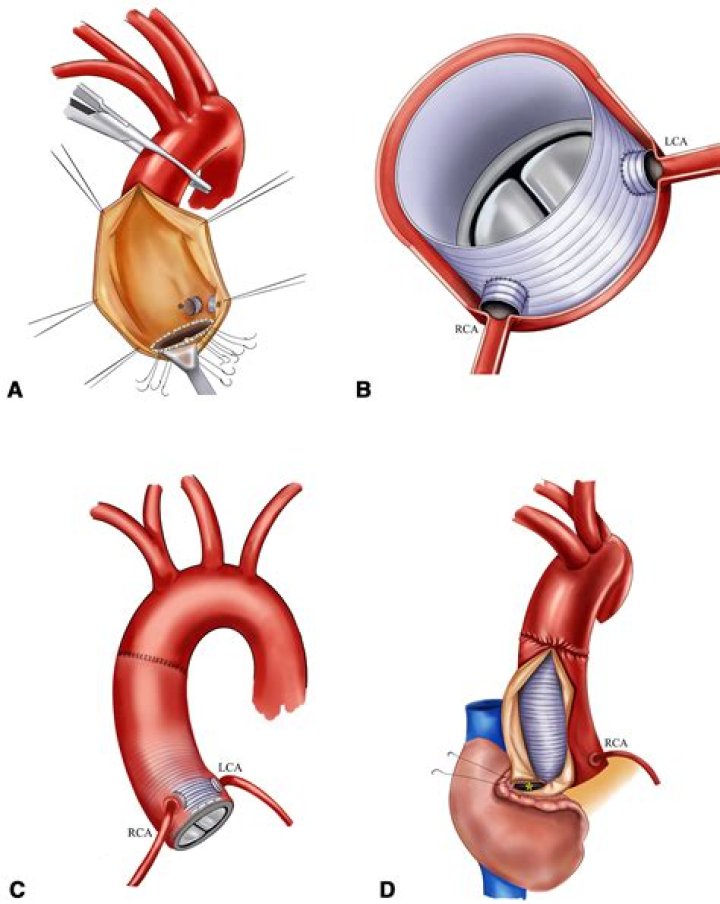
The original Cabrol technique comprises of ascending aortic replacement with a composite graft and direct reimplantation of coronary arteries into an interposition graft, which is anastomosed side-to-side to the aortic graft [1].
What is the ascending aorta and arch?
The ascending aorta begins at the heart’s left ventricle and extends to the aortic arch, or the bend in the aorta. The arch of the aorta gives off branches to the head and arms.
Which complaint is most commonly associated with an ruptured aortic arch aneurysm?
Symptoms of a thoracic aortic aneurysm are most evident when the aneurysm occurs where the aorta curves down (aortic arch). They may include: Chest pain, generally described as deep and aching or throbbing. This is the most frequent symptom.
What causes the ascending aorta to dilate?
The diameter at the level of the ascending aorta measures 4.6 cm. The diameter of the aortic root is normal….Table 1.
| Diameter | Value | Study |
|---|---|---|
| Proximal ascending aorta, cm | ||
| Men | 2.9±0.3 | TTE |
| Women | 2.6±0.3 | TTE |
| Ascending aorta, cm/m2 | 1.4–2.1 cm/m2 | TEE |
What is a Cabrol graft?
The Cabrol technique is a surgical technique for reconstruction of coronary arteries after aortic root replacement. It uses a Dacron graft interposed between the aortic root graft and native coronary artery [1].
What is the aortic arch?
The aortic arch is the top part of the main artery carrying blood away from the heart. Aortic arch syndrome refers to a group of signs and symptoms associated with structural problems in the arteries that branch off the aortic arch.
Where is the ascending aortic arch?
Your Ascending Aorta and Aortic Arch The ascending aorta begins above the aortic root and extends towards the neck until it begins to turn and give rise to the aortic arch. The ascending aorta is more frequently affected by aneurysms and dissections and requires open heart surgery to be repaired.
What is LOEY Dietz syndrome?
Loeys-Dietz syndrome is a connective tissue disorder that was first described in 2005. Most individuals with this disorder have craniofacial features that include hypertelorism (widely spaced eyes) and a bifid or broad uvula.
How serious is a mildly dilated ascending aorta?
Such dilatation of the ascending aorta frequently leads to significant aortic valvular insufficiency, even in the presence of an otherwise normal valve. The dilated or aneurysmal ascending aorta is at risk for spontaneous rupture or dissection.
How common is a dilated ascending aorta?
According to the CDC, the incidence of ascending TAA is estimated to be around 10 per 100,000 person-years. Women and men have similar incidences of thoracic aortic aneurysm but the age at diagnosis is a decade higher in women (70s) than in men (60s).
What are the 6 aortic arches?
| Arch | Vessel |
|---|---|
| 4th Aortic sac | R: Right subclavian artery L: Aortic arch Brachiocephalic artery (divides into right subclavian and right common carotid artery) |
| 5th | R: Right pulmonary artery (proximal part) L: Ductus arteriosus |
| 6th Intersegmental artery | Left pulmonary artery |
Where is the aortic arch located in the heart?
Location of the Aortic Arch. The aortic arch is the part of the aorta between the ascending aorta and thoracic descending aorta. The sharpness of the angle can be different among individuals.
Can the angle of the aortic arch cause dissection?
In some people, the angle of the aortic arch coupled with certain medical conditions can lead to aortic dissection where the ascending aorta meets the aortic arch. Aortic dissection occurs when a tear in the tunica intima allows blood to be pushed between the tunica intima and the tunica media.
What is right aortic arch with aberrant left subclavian artery?
Right aortic arch with aberrant left subclavian artery is the most common of the several types of right aortic arch, occurring in 0.05% to 0.1% of the population. 5 It results from involution of the left aortic arch between the left common and left subclavian arteries with persistence of the embryologic right arch.
How is the aortic arch connected to the pulmonary trunk?
The arch is still connected to the pulmonary trunk by the ligamentum arteriosum (remnant of the foetal ductus arteriosus). There are three major branches arising from the aortic arch. Proximal to distal: Brachiocephalic trunk: The first and largest branch that ascends laterally to split into the right common carotid and right subclavian arteries.